

New PhD!! - When and where will the tumor return? New findings may hold the answer
This article presents the doctoral research of Marianne Hjellvik Hannisdal, conducted at the University of Bergen, which investigates how advanced analysis of medical imaging and artificial intelligence can improve understanding of glioblastoma progression. By exploring patterns of tumour growth, recurrence, and treatment response, the work aims to identify predictive markers that may contribute to more precise, individualised clinical management of this highly aggressive brain tumour.
Published: (Updated: )

Among malignant brain tumours in adults, glioblastoma is the most frequent, aggressive and incurable form of grade 4 astrocytoma. Despite advanced treatment combining surgery, radiotherapy and chemotherapy, survival remains dismal, often only 9–12 months.
One of the main reasons for this poor prognosis is the complexity of the tumour. Glioblastoma is not a single uniform disease, but rather a collection of many different subtypes. There is considerable variation both within a single tumour and between patients, in terms of cellular composition, genetic alterations and biological behaviour. This heterogeneity directly affects how the tumour responds to treatment.
In addition, glioblastoma grows in a diffuse and infiltrative manner. Individual tumour cells migrate away from the main tumour mass and into surrounding brain tissue—often beyond what can be detected with conventional imaging. These cells may remain outside treatment margins, giving rise to new tumour growth and recurrence. This infiltrative behaviour is a defining hallmark of the disease and a key reason why relapse is almost inevitable.
Despite this complexity, most patients today receive largely the same standard treatment, with limited adaptation to their individual tumour biology. This may lead to suboptimal treatment for some patients. At the same time, clinical assessment of tumour extent relies heavily on visual interpretation of MRI scans, which can vary between experts.
But what if we could see beyond what the eye can detect?
What if we could map tumour extent more precisely—capturing not only the clearly visible regions, but also the more diffuse and difficult-to-detect components? And what if, already at the time of diagnosis, we could predict where in the brain a tumour is likely to recur, and when?
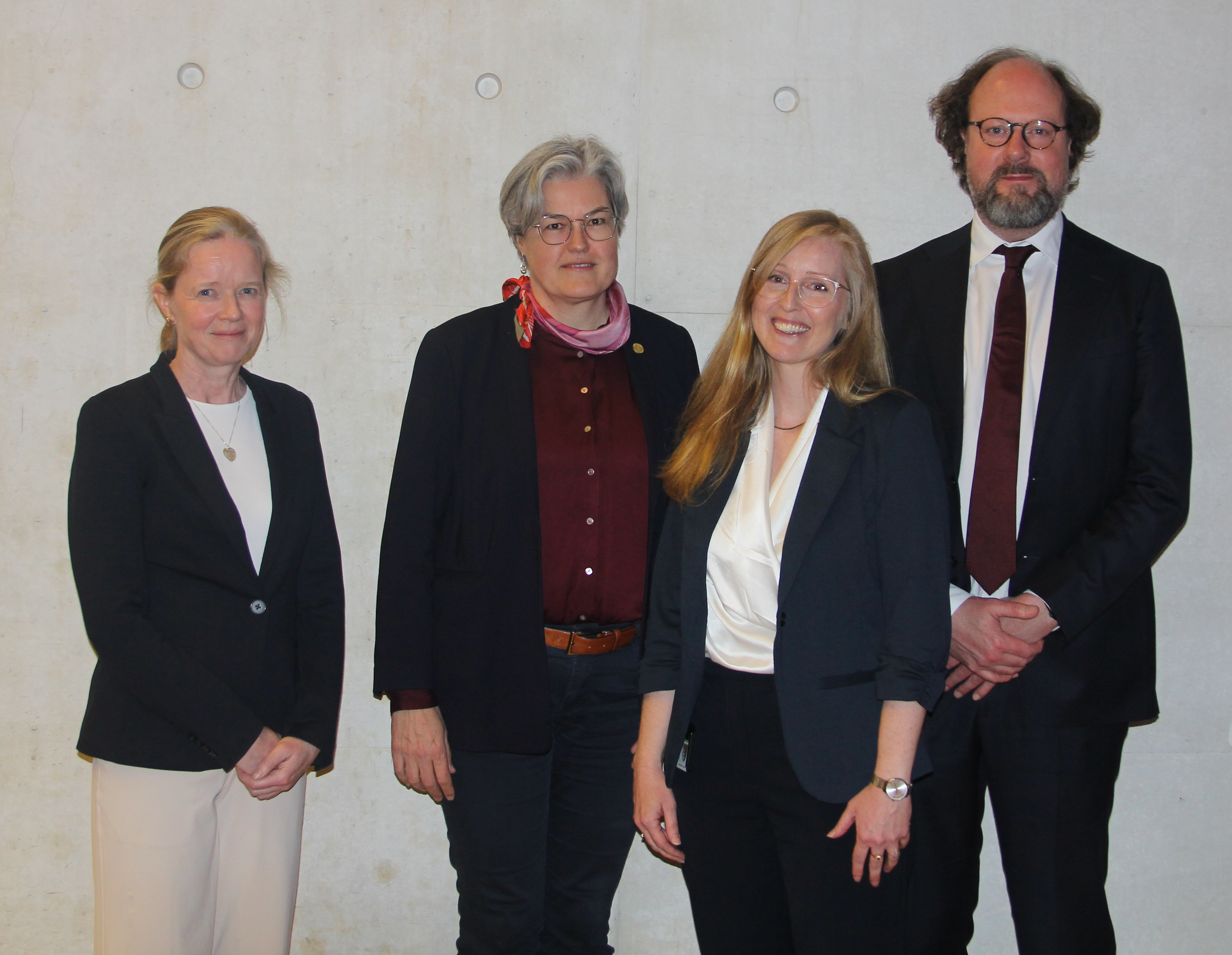
These are the questions explored by Marianne Hjellvik Hannisdal in her doctoral work at the University of Bergen.
In her research, she used artificial intelligence based on deep learning and machine learning to analyse multiparametric MRI data. The models were applied to quantify both visible and less visible tumour volumes, and the results were compared with manual delineations performed by experienced clinicians.
The findings showed that the relationship between the contrast-enhancing (visible) part of the tumour and the non-enhancing (less visible) component has prognostic value. This ratio was associated with both the location of tumour recurrence in the brain and the time to recurrence. Importantly, these relationships were influenced by patient-specific factors such as age and tumour genetics.
In other words, it may be possible to identify meaningful patterns already at the time of diagnosis that provide insight into how and where the disease will progress.
This opens new opportunities for how glioblastoma is treated. Instead of relying on standardised treatment margins based on historical data, future strategies could be tailored to the individual patient. Some patients may benefit from narrower treatment margins, while others may require more intensive therapy, depending on their biological profile.
These findings may also influence how patients are monitored. By identifying brain regions at higher risk of recurrence, follow-up imaging and surveillance can be more precisely targeted.
Finally, the study demonstrates how combining imaging data with genetic information can help identify biomarkers that explain differences in treatment response. Patients may thus be stratified into subgroups with distinct disease trajectories and survival outcomes.
Taken together, this work represents an important step towards more personalised treatment of glioblastoma—with the ultimate goal of improving outcomes and quality of life for patients.
References
1. Deep learning prediction of features underlying spatiotemporal tumour recurrence patterns as indicators of treatment response in grade 4 glioma, Hannisdal MH et al, Preprint 2025: DOI: 10.2139/ssrn.5177606 (external link) (Under review after revision for Nature Precision oncology
2. Exploiting Deep Learning to Enhance Tumour-conformed Delineation and Reduced Isotropic Margin in Radiotherapy: Updated ESTRO-EANO Guidelines. (external link) Hannisdal MH et al., Clin Oncol (R Coll Radiol). 2023 Oct;35(10
3. Feasibility of deep learning-based tumor segmentation for target delineation and response assessment in grade-4 glioma using multi-parametric MRI. (external link) Hannisdal MH et al., Neurooncol Adv. 2023 13;5(1):vdad037.
We are grateful to Samarbeidsorganet HelseVest that financed her PhD position, Kreftforeningen and KLINBEFORSK for financial support for our national phase IB/II BORTEM-17 study (NCT03643549) and the Brain Tumour Society´s members for user involvement.
